Research Article | | Peer-Reviewed

Efficacy of Antifungal Medicines Against Clinical Isolates of Candida Species Responsible for Vaginal Candidiasis from Regional Referral Hospitals in Tanzania

Received: 20 October 2025     Accepted: 29 October 2025     Published: 9 December 2025
Views:       Downloads:
Abstract

Vaginal candidiasis, caused by Candida species affects a significant proportion of women worldwide. Although different antifungal medicines are being used to treat the infection, the emergence of resistance against these medicines have been reported. This study assessed the efficacy of antifungal medicines which are recommended for treatment in the Tanzania Standard Treatment Guidelines against clinical isolates of Candida spp. Presumptive Isolates of Candida spp. collected from Regional Referral Hospitals (RRH) were transported to the Tanzania Medicine and Medical Devices Authority (TMDA) Microbiology laboratory for identification and confirmation by PCR. The confirmed isolates were used for susceptibility testing to establish the efficacy of antifungal medicines. Results indicate that Candida albicans (54%) was the most prevalent Candida spp. among isolates collected. Among the antifungal medicines tested, Miconazole showed the highest efficacy against 229 isolates of Candida spp. with 89.2% of the isolates being susceptible. This was followed by Clotrimazole, with 79.9% and Nystatin, 79.5% of isolates were susceptible, while Voriconazole showed moderate efficacy with 68.9%. Fluconazole demonstrated the lowest efficacy among the tested antifungals with 63.3%. Both Candida albicans and non-albicans Candida spp. displayed similar susceptibility patterns. Out of the 229 Candida spp. isolates tested, 4.4% were found to exhibit multidrug resistance. Findings from this study suggest that despite the observed resistance, the majority of antifungal medicines listed in the Standard Treatment Guidelines in Tanzania remain effective and are still considered reliable for treating vaginal candidiasis.

Published in American Journal of Life Sciences (Volume 13, Issue 6)
DOI 10.11648/j.ajls.20251306.13
Page(s) 198-207
Creative Commons

This is an Open Access article, distributed under the terms of the Creative Commons Attribution 4.0 International License (http://creativecommons.org/licenses/by/4.0/), which permits unrestricted use, distribution and reproduction in any medium or format, provided the original work is properly cited.

Copyright

Copyright © The Author(s), 2025. Published by Science Publishing Group

Keywords

Antifungal Efficacy, Candida albicans, Vaginal Candidiasis, Antifungal Resistance, Multidrug Resistance

1. Introduction
Vaginal candidiasis, commonly referred to as a yeast infection, is a prevalent fungal infection affecting the mucosal surfaces of female genital tract . The primary causative agent of vaginal candidiasis is Candida albicans, a commensal microorganism that reside in the gastrointestinal, respiratory, and genitourinary tracts . It has been reported that C. albicans constitutes approximately 80.5% of the cases of vaginal candidiasis, with other Candida spp. such as Candida glabrata also plays a significant role .
Globally, vaginal candidiasis affects a significant proportion of women, causing considerable discomfort and disruption to daily activities. Studies in various countries have shown a high prevalence of this infection, highlighting its significant impact on women's health . In Africa, the burden of vaginal candidiasis is similarly substantial, with many women experiencing recurrent infections that affect their quality of life . Factors such as limited access to healthcare, lack of awareness, and varying socioeconomic conditions contribute to the challenges in managing this infection across the continent . Similarly, in Tanzania, vaginal candidiasis remains a common health issue among women, although comprehensive data on its prevalence and the antifungal susceptibility of isolates of Candida spp. are limited. This lack of detailed epidemiological information hampers the development of effective treatment strategies and the management of antifungal resistance in the region .
Vaginal candidiasis is characterized by symptoms such as itching, burning, soreness, and abnormal vaginal discharge . This infection affects a significant proportion of women at some point in their lives. Several risk factors have been reported to contribute to the development of vaginal candidiasis, including the prolonged use of antibiotics, diabetes mellitus, pregnancy, immunodeficiency, use of oral contraceptives, sexual activity, and certain hygiene practices . These factors can disrupt the normal vaginal microflora, creating an environment conducive to Candida overgrowth and infection . The management of vaginal candidiasis typically involves antifungal therapy. Fluconazole, which is an azole antifungal has been commonly used for treatment; however, the emergence of Fluconazole-resistant strains of Candida has been reported, posing a significant challenge to effective management . Other antifungal agents such as Clotrimazole, Miconazole, Nystatin, and Terbinafine are also employed, with varying degrees of efficacy against different Candida spp. . Given the increasing incidence of antifungal resistance, there is a pressing need to evaluate the susceptibility of clinical isolates of Candida spp. to commonly used antifungal medicines . This is particularly important in countries like Tanzania, where data on the prevalence and antifungal susceptibility of isolates responsible for vaginal candidiasis is limited. Understanding the local epidemiology and resistance patterns of Candida spp. will aid in developing targeted treatment strategies and improve patient outcomes.
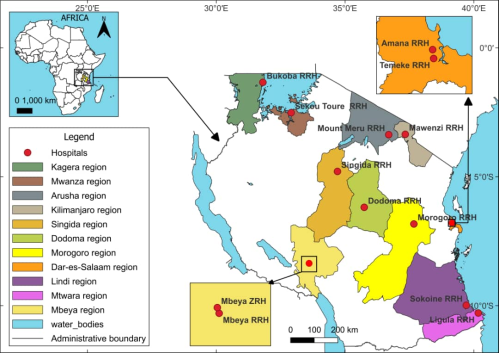
Figure 1. Map of Tanzania showing coverage of regions for sample collection.
The prevalence of vaginal candidiasis among women in Tanzania underscore the importance of ensuring that the treatments prescribed are effective in eliminating the infection and preventing recurrence . Antifungal resistance is a growing global concern, and Tanzania is not exempted from this challenge. TMDA is tasked with function of ensuring that medicines used in Tanzania are of acceptable quality, safe and efficacious. To accomplish this role, TMDA utilizes different strategies such as medicines registration, inspection, post-market surveillance, pharmacovigilance and conducting various researches. Therefore, the objective of this study is to assess the efficacy of antifungal medicines listed in the Standard Treatment Guidelines against clinical isolates of Candida species from selected regional referral hospitals in Tanzania.
2. Methodology
2.1. Study Area
The study was conducted between September 2024 and June 2025 in Tanzania mainland, focusing on selected regional referral and zonal hospitals equipped with functional microbiological laboratories that perform microbial cultures as part of their diagnostic procedures.
2.2. Study Design and Sample Size Determination
A cross-sectional study design was employed, utilizing a purposive (selective) sampling technique. Only isolates confirmed to be Candida spp. obtained from various regional referral hospitals in Tanzania mainland were included in the study. TMDA issued official letters to eligible hospitals, requesting the isolates for use in the study. The targeted number was 30 isolates of Candida spp. per hospital.
2.3. Sample Collection
The study focused on previously isolated and stored Candida spp. from vaginal swabs of patients diagnosed with vaginal candidiasis in the selected hospitals. TMDA laboratory analyst collected isolates from all the selected hospitals, transported them in transport media in cool temperature within 24 hours to the TMDA Microbiology Laboratory located in Dar es Salaam for subsequent analyses.
2.4. Laboratory Methods
2.4.1. Isolation of Candida spp.
Isolates were pre-enriched with Sabouraud Dextrose Broth (SDB) (Oxoid UK) at 32.5°C for 24 hours, followed by subculturing using a streaking method on primary media, Sabouraud Dextrose Agar (SDA) (Oxoid UK) at 32.5°C for 24 to 48 hours. Presumptive Candida spp. colonies on SDA were smooth, creamy, and white to off-white in colour, soft or slightly pasty or mucoid texture, and medium to large size with well-defined edges. Gram staining was performed to observe the yeast cells.
2.4.2. Identification Test for C. albicans by Germ Tube Test
A germ tube test was performed to differentiate C. albicans and non-albicans Candida spp. the presumptive isolates were suspended in 1mL of human serum and incubated at 37 °C for 2 hours. After incubation, a drop of suspension was mounted on microscopic slides, covered and observed under a microscope ×10 objective for germ tube formation as previously reported by DN Anh. et al .
2.4.3. DNA Extraction and Molecular Detection by PCR
DNA was extracted from Candida spp. isolates identified by using the Fungi/Yeast Genomic DNA Isolation Kit (Norgen Biotek Corp.), following the manufacturer’s instructions as described by Kumar & Mugunthan (2018) for enhancing DNA yield and purity. The molecular identification of C. albicans was achieved through the amplification of a specific region within the CALB gene, which encodes an enzyme involved in the hydrolysis of β-glucosides in carbohydrates, as previously described by F. Mallus et. al . The primers utilized for this amplification were CalB-F (5'-TTTATCAACTTGTCACACCAGA-3') and CalB-R (5'-ATCCCGCCTTACCACTACCG-3') (Inqaba Biotechnical Industries (Pty) Ltd, South Africa) with base pairs 273 was used. The polymerase chain reaction was performed PCR instrument Surecycler 8800 (Agilent Technologies) and conditions as described by Mallus et al. . The reaction mixture contained 3 µL aliquot of the extracted DNA in 10 mM Tris and 1 mM EDTA (TE buffer), 12.5 µL of the master mix, 0.5 µL each of the forward and reverse primers, and 7.5 µL of nuclease-free water to reach the final volume of 25 µL. The PCR amplification conditions were as follows: Initial step heating at 94°C for 15 seconds, followed by 40 cycles of 94°C for 30 seconds, 63°C for 1.5 minutes, and 72°C for 1.5 minutes, with a final extension step at 72°C for 10 minutes, yielding an expected amplicon size of 273 base pairs. A 5 µL PCR product was stained with 0.5 μg/mL Ethidium bromide and loaded into 1% agarose gel submerged in 1x Tris-acetate-EDTA (TAE) buffer for gel electrophoresis in a Biometra electrophoresis tank (Analytik Jena Company) for 45 minutes. The PCR product was visualized under UV light using a benchtop Trans Illuminator (BioDoc It TM) and images was captured.
2.4.4. Antifungal Susceptibility Testing
The antifungal susceptibility of isolated C. albicans was evaluated as described in the CLSI guidelines M27M44S, third edition (2022) . Suspensions of overnight cultures were prepared in sterile phosphate-buffered saline (Glentham Life Sciences GX 8368). Turbidity was adjusted to a 0.5 McFarland standard using a calibrated UV-spectrophotometer (Thermo Scientific Helios Zeta UV-VIS-USA), resulting in an inoculum containing 1-5×10⁶ CFU/mL. A 25µL. aliquot of the suspension was inoculated onto Mueller-Hinton agar (Sigma Adrich Lot BCCD 3626) supplemented with 2% glucose and 0.5µl/mL methylene blue dye and allowed to dry for 15 minutes at room temperature in a laminar air flow cabinet. Antifungal discs of Fluconazole 25µg (Biomaxima Lot 420/06/Z), Clotrimazole 10 µg (Himedia Lot 0000614732), Miconazole 30µg (Himedia Lot 620845), Voriconazole 1µg (Himedia Lot000643162) and Nystatin100 units (Oxoid U. K) were placed on the inoculated agar plates using sterile forceps. The plates were incubated at 35 ± 2°C for 24 hours in calibrated incubator (Memmert loding model 30-1060). After incubation time, the diameters of the growth inhibition zone were measured manually using a calibrated Venier caliper. The results were interpreted according to CLSI guidelines M27M44S, third edition (2022) as either sensitive, resistant, or intermediate. C. albicans ATCC 10231 was used as a positive standard in all tests requiring a positive control.
2.5. Data Analysis
Data from laboratory tests were compiled in Microsoft Excel and analyzed using Statistical Product and Service Solutions (SPSS) V20 (IBM USA). Descriptive statistics were used to summarize the frequencies of Candida spp. and their antifungal resistance patterns. A bivariate analysis using the Chi-square test was performed to assess variations in Candida spp. distribution and antifungal resistance patterns across hospitals, and the P-value <0.05 was considered statistically significant.
3. Results and Discussion
3.1. Sample Size and Response Rate
A total of 242 Isolates were collected from all selected hospitals. The collected isolates registered a response rate of 63% of the expected sample size (N=384) from all 14 hospitals. The expected sample size per hospital was at least 30 isolates. However, the actual number of isolates per hospital varied due to the local prevalence of Candida spp.
3.2. Isolation and Identification of Candida spp.
Among the collected isolates (N=242), 94.6% (n=229) were confirmed as Candida spp. based on the morphological characteristics when cultured on SDA plates. Colonies were observed to be creamy, smooth, and circular. The microscopical examination of the germ tube test and Gram staining as shown in Figure 3. Indicated that 52.4% (n=120) were C. albicans, while Candida spp. altogether were 47.6% (n=109). This study found that 5.4% (n=13) of the total hospital reported cases of Candida spp. were not Candida spp. suggesting diagnosis error.
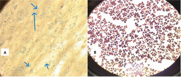
Figure 2. Microscopic identification of Candida spp. using wet mount image (A) and Gram staining image (B).
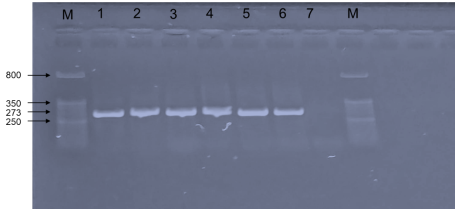
Figure 3. Gel electrophoresis image of PCR product of CalB gene (273 bp) for identification of C. albicans, whereby Lane M marker, Lane 1 Standard C. albicans ATCC 10231 (positive control) Lane 2-6 positive samples of C. albicans isolates, Lane 7 negative control.
3.3. Confirmation of C. albicans
Confirmation for C. albicans were performed by PCR on 229 samples collected from 14 Regional Referral Hospitals as shown in Figure 3. Results of PCR confirmed that 52.4% (n= 120) of the isolates were C. albicans, while 47.6% (n=109) were non albicans Candida spp.
3.4. Prevalence of C. albicans Across Regional Referral Hospitals
The prevalence of C. albicans varied across hospitals. Sekou Toure RRH had the highest prevalence with 100% (n=5) of isolates identified as C. albicans, followed by Amana RRH at 83.3% (n=6), Morogoro RRH at 82.6% (n=23), and Dodoma RRH at 66.7% (n=23). Other hospitals with high prevalence included Bukoba RRH 64.0% (n=16) and Singida RRH 61.1% (n=11). In contrast, the lowest prevalence was observed at Mwananyamala RRH 0% (n=0), Mbeya Zonal Hospital 11.1% (n=9) and Mount Meru RRH 15% (n=3) as represented in Table 1. There was a statistically significant variation in C. albicans prevalence across hospitals (χ² = 44.47, p value = 0.001), in such that Sekou Toure and Amana RRH showed the highest proportions.
Overall, the findings confirm that C. albicans was more prevalent among the Candida spp. isolates from patients with candidiasis, with notable variation in prevalence across different healthcare facilities. A similar prevalence has been reported in other studies . However, findings from this study indicated that there is no serious gap between the prevalence of C. albicans and other Candida spp. Hence, this study suggests that other Candida spp. are gaining more ground on the prevalence scale, contrary to previous reported cases .
Table 1. Comparison of C. albicans and Non-albicans Candida spp. prevalence across hospitals using Chi-Square Test (N=229).

Hospital Name

Prevalence of Candida spp.

X2

p - value

C. albicans n (%)

Non-albicans Candida spp. n (%)

Amana RRH

5 (83.3)

1 (16.7)

44.468

<0.001

Bukoba RRH

16 (64)

9 (36)

Dodoma RRH

22 (66.7)

11 (33.3)

Ligula RRH

12 (44.4)

15 (55.6)

Mawenzi RRH

3 (42.9)

4 (57.1)

Mbeya RRH

8 (53.3)

7 (46.7)

Mbeya Zonal

1 (11.1)

8 (88.9)

Morogoro RRH

19 (82.6)

4 (17.4)

Mount Meru RRH

3 (15)

17 (85)

Mwananyamala RRH

0 (0)

2 (100)

Sekou Toure RRH

5 (100)

0 (0)

Singida RHH

11 (61.1)

7 (38.9)

Sokoine RRH

12 (35.3)

22 (64.7)

Temeke RRH

3 (60)

2 (40)

Total

120 (52.4)

109 (47.6)

3.5. Antifungal Susceptibility Test by Disc Diffusion
The clear zones of inhibition observed surrounding the antifungal disks indicated the effectiveness of the respective antifungal agent against the Candida spp. isolates. The larger the zones the higher the susceptibility. Meanwhile, the smaller or no clear zones suggest resistance when the same antifungal is tested against all isolates. The varying sizes of the inhibition zones demonstrate differences in susceptibility patterns of the isolates to the tested antifungal agents. Results from this study were found to be in the zone limits for resistant, intermediate and susceptible as prescribed in the CLSI guidelines M27M44S, third edition (2022. This suggest that the obtained results are scientifically acceptable.
3.6. Antifungal Susceptibility Profile of Candida spp. Isolates (All Isolates Combined)
The antifungal susceptibility of Candida spp. isolates (N=229) was assessed against five commonly used antifungal agents, out of which Miconazole showed the highest efficacy, with 89.2% (n = 204) of isolates classified as susceptible, 6.1% (n = 14) displayed resistance, while 4.9% (n=11) were intermediately resistant. Similarly, Clotrimazole and Nystatin demonstrated good performance, with 79.9% (n = 183) and 79.5% (n = 182) of isolates susceptible, respectively. Voriconazole showed moderate efficacy, with 68.9% (n = 156) of isolates susceptible. Fluconazole demonstrated the lowest efficacy among the tested antifungals, with 63.3% (n = 145) of isolates classified as susceptible and 31.0% (n = 71) resistant while 5.1 (n=13) were intermediate. This indicates a concern about the possibility of reduced efficacy, as shown in Table 2. These results suggest that while most Candida spp. isolates remain susceptible to azoles and polyenes, there is an observable trend of increased resistance, particularly to Fluconazole and Voriconazole. Findings from similar studies elsewhere also agree that resistance toward fluconazole is a growing concern, hence the need for monitoring. Surveillance of antifungal drug resistance displayed dynamism and inconsistency between institutions and countries . This report from a localized study in Tanzania contributes to the global picture of emerging resistance toward antifungal drugs and hence supports the need for stern action.
Table 2. Susceptibility Distribution of Candida spp. Isolates (N=229) to antifungal medicine recommended in the Standard Treatment Guideline.

Antifungal agent

Susceptible (S) n (%)

Intermediate (I) n (%)

Resistant (R) n (%)

Miconazole (MIC)

204 (89.19)

11 (4.9)

14 (6.1)

Voriconazole (VRC)

156 (68.9)

4 (1.7)

69 (30.1)

Clotrimazole (CC)

183 (79.9)

16 (7)

30 (13.1)

Fluconazole (FLC)

145 (63.3)

13 (5.7)

71 (31)

Nystatin (NS)

182 (79.5)

6 (2.6)

41(17.9)

3.7. Antifungal Susceptibility Profile of C. albicans
The highest susceptibility of C. albicans was observed with Miconazole at 86.7%(n=104), followed by Nystatin at 84.2% (101) and Clotrimazole at 80.8% (97). In contrast, Voriconazole and Fluconazole exhibited relatively higher resistance rates of 29.2% (n=35) and 30.8% (n=37), respectively, as shown in Figure 4 below. These findings suggest that while most Candida albicans isolates remain susceptible to azoles and polyenes, there is a growing concern of resistance, particularly to Fluconazole and Voriconazole, indicating the need for continuous antifungal resistance monitoring and prudent antifungal usage. This study suggests that, given the observed emerging resistance to Fluconazole among C. albicans isolates, clinicians may need to prioritize agents like Miconazole and Nystatin, which demonstrated >80% efficacy. Although similar trends have been observed elsewhere , the current study showed varied susceptibility such that both C. albicans and non albicans Candida spp. present a similar susceptibility profile. This phenomenon suggests for establishment of phylogenetic relationships among species and possibly the responsible resistant genes.
Given the significant resistance to Fluconazole among C. albicans isolates, clinicians may need to prioritize agents like Miconazole and Nystatin, which demonstrated >80% efficacy.
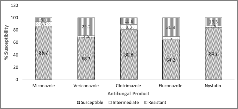
Figure 4. Susceptibility Profile of Candida albicans Isolates to Commonly Used Antifungals.
3.8. Antifungal Susceptibility Profile of Non-albicans Candida spp.
The antifungal susceptibility profile of Candida spp. isolates tested against five commonly used antifungal agents: Miconazole, Voriconazole, Clotrimazole, Fluconazole, and Nystatin. The results indicate that Miconazole exhibited the highest susceptibility rate with 91.7% (n=100) of isolates being sensitive, followed by Clotrimazole 78.9% (n=86) and Nystatin 74.3% (81). However, Voriconazole and Fluconazole demonstrated higher resistance rates of 31.2% (n=34) each, as shown in Table 3. These findings suggest that while most Candida spp. isolates were still susceptible to commonly used antifungals, the emerging concern of resistance, particularly towards Fluconazole and Voriconazole, highlighting the need for continuous antifungal resistance monitoring and rational drug use in clinical settings. Similar patterns have been reported elsewhere and these rise concerns about the need for a wider global picture of the situation.
Table 3. Antifungal Susceptibility Profile on non- albicans Candida spp. Isolates (N=109).

Drug

Susceptible n (%)

Intermediate n (%)

Resistant n (%)

Miconazole

100 (91.7)

3 (2.8)

6 (5.5)

Voriconazole

74 (67.9)

1 (0.9)

34 (31.2)

Clotrimazole

86 (78.9)

6 (5.5)

17 (15.6)

Fluconazole

68 (62.4)

7 (6.4)

34 (31.2)

Nystatin

81 (74.3)

3 (2.8)

25 (22.9)

3.9. Prevalence of Multi-drug Resistance (MDR) Among Candida spp. Isolates
The MDR results indicate that out of the Candida spp. isolates (N=229) analyzed, 4.4% (n=10) of isolates were found to exhibit multidrug resistance (MDR) defined as resistance to two or more classes of antifungal agents. The remaining 95.6% (n=219) of isolates were classified as non-Multi Drug Resistant (Non-MDR). The distribution of MDR cases across the participating hospitals (N=14) is presented in Table 4. While the majority of hospitals reported non-MDR cases, a few facilities (n=4) showed a presence of multidrug-resistant isolates. Notably, among the isolates, Ligula RRH N=27 reported the highest MDR prevalence at 14.8% (n = 4), followed by isolates from Bukoba RRH (N=25) at 12.0% (n=3), and isolates from Mount Meru RRH (N=20) at 10.0% (n=2) and isolates from Mbeya RRH (N=15) reported MDR of 6.7% (n=1). The rest of the hospitals recorded no MDR isolates.
There was no statistically significant difference in MDR occurrence between hospitals (X² = 14.842, p value = 0.129) as illustrated in Table 4 indicating that the observed differences may be due to random variation. These findings suggest that, although overall MDR prevalence was low (4.4%), some hospitals, Ligula RRH, Bukoba RRH and Mount Meru RRH showed disproportionately higher rates. While not statistically significant (p-value = 0.129), these sites may benefit from targeted surveillance due to potential clustering. Although several studies have reported on the resistance towards antifungal medicines , the current study highlighted the possibility of emerging multidrug resistance, a case also highlighted in other reports . The observed phenomena may need to be studied in different global regions to contribute to the recent global picture of the situation.
Table 4. Prevalence of Multidrug Resistance (MDR) Among Candida spp. Isolates Across Hospitals.

Hospital Name

MDR Status

X2

p - value

MDR n (%)

Not MDR n (%)

Amana RRH

0 (0)

6 (100)

14.842

0.129

Bukoba RRH

3 (12)

22 (88)

Dodoma RRH

0 (0)

33 (100)

Ligula RRH

4 (14.8)

23 (85.2)

Mawenzi RRH

0 (0)

7 (100)

Mbeya RRH

1 (6.7)

14 (93.3)

Mbeya Zonal

0 (0)

9 (100)

Morogoro RRH

0 (0)

23 (100)

Mount Meru RRH

2 (10)

18 (90)

Mwananyamala RRH

0 (0)

2 (100)

Sekou Toure RRH

0 (0)

5 (100)

Singida RHH

0 (0)

18 (100)

Sokoine RRH

0 (0)

34 (100)

Temeke RRH

0 (0)

5 (100)

Total

10 (4.4)

219 (95.6)

4. Conclusion
This study generally demonstrated that Candida albicans were slightly more prevalent than other Candida spp. among the isolates collected from selected regional referral hospitals in Tanzania. However, a significant variation in prevalence was observed across hospitals. Antifungal susceptibility testing revealed a good efficacy of Miconazole, Clotrimazole, and Nystatin while resistance was observed in Fluconazole and Voriconazole. The prevalence of multidrug-resistant Candida spp. was low, but isolated hotspots were identified. Despite the observed emerging resistance, the majority of antifungals listed in the Standard Treatment Guideline are efficacious, hence still reliable for the treatment of vaginal candidiasis against all Candida spp. Findings from this study recommend strengthening continuous surveillance of Candida species and their antifungal susceptibility profiles across healthcare facilities in Tanzania.
Abbreviations

ATCC

American Type Culture Collection

CLSI

Clinical and Laboratory Standards Institute

MDR

Multi Drug Resistance

PCR

Polymerase Chain Reaction

RRH

Regional Referral Hospital

SDA

Sabouraud Dextrose Agar

SDB

Sabouraud Dextrose Broth

SPP

Species

SPSS

Statistical Product and Service Solutions

TAE

Tris-acetate-EDTA.

TMDA

Tanzania Medicines and Medical Devices Authority

Acknowledgments
We would like to acknowledge the contribution of Mr. John Mwingira, who supported the statistical analysis of the data from this study to produce scientifically meaningful results.
Author Contributions
Adelard Bartholomew Mtenga: Conceptualization, Data curation, Formal Analysis, Methodology, Supervision, Writing – original draft, Writing – review & editing
Adam Mitangu Fimbo: Funding acquisition, Project administration, Resources, Supervision, Writing – review & editing
Danstan Hipolite Shewiyo: Formal Analysis, Project administration, Supervision, Writing – review & editing
Saxon Joseph Mwambene: Data curation, Investigation, Methodology, Writing – review & editing
Revocatus Evarist Makonope: Data curation, Investigation, Methodology, Writing – original draft
Shaban Bikiz Kombo: Data curation, Investigation, Methodology
Funding
This study did not receive any funds from an external source.
Data Availability Statement
The data supporting the outcome of this research work has been reported in this manuscript.
Conflicts of Interest
Authors declares no conflict of interest.
References
[1] S. W. Kashem et al., “Candida albicans morphology and dendritic cell subsets determine T helper cell differentiation,” Immunity, vol. 42, no. 2, pp. 356–366, 2015,
[2] S. S. Richter, R. P. Galask, S. A. Messer, R. J. Hollis, D. J. Diekema, and M. A. Pfaller, “Antifungal susceptibilities of Candida species causing vulvovaginitis and epidemiology of recurrent cases,” J. Clin. Microbiol., vol. 43, no. 5, pp. 2155–2162, 2005,
[3] W. Lin, M. Karin, W. Lin, and M. Karin, “Review series A cytokine-mediated link between innate immunity, inflammation, and cancer,” vol. 117, no. 5, pp. 1175–1183, 2007,
[4] J. M. Achkar and B. C. Fries, “Candida infections of the genitourinary tract,” Clin. Microbiol. Rev., vol. 23, no. 2, pp. 253–273, 2010,
[5] D. N. Anh et al., “Prevalence, species distribution and antifungal susceptibility of Candida albicans causing vaginal discharge among symptomatic non-pregnant women of reproductive age at a tertiary care hospital, Vietnam,” BMC Infect. Dis., vol. 21, no. 1, pp. 1–10, 2021,
[6] C. Chayachinda, T. Rekhawasin, M. Thamkhantho, and P. Aneklap, “Acute Vaginal Candidiasis: A review of treatment guidelines and Siriraj experience,” Thai J. Obstet. Gynaecol., vol. 29, no. 6, pp. 306–312, 2021,
[7] A. O. Mohamed et al., “Prevalence of vulvovaginal candidiasis among pregnant women in Africa: A systematic review and meta-Analysis,” J. Infect. Dev. Ctries., vol. 16, no. 8, pp. 1243–1251, 2022,
[8] B. Gonçalves, C. Ferreira, C. T. Alves, M. Henriques, J. Azeredo, and S. Silva, “Vulvovaginal candidiasis: Epidemiology, microbiology and risk factors,” Crit. Rev. Microbiol., vol. 42, no. 6, pp. 905–927, 2016,
[9] M. F. Mushi, O. Bader, C. Bii, U. Groß, and S. E. Mshana, “Virulence and susceptibility patterns of clinical Candida spp. isolates from a tertiary hospital, Tanzania,” Med. Mycol., vol. 57, no. 5, pp. 566–572, Jul. 2019,
[10] J. D. Sobel, “Recurrent vulvovaginal candidiasis,” Am. J. Obstet. Gynecol., vol. 214, no. 1, pp. 15–21, 2016,
[11] J. de Cássia Orlandi Sardi et al., “Vulvovaginal Candidiasis: Epidemiology and Risk Factors, Pathogenesis, Resistance, and New Therapeutic Options,” Curr. Fungal Infect. Rep., vol. 15, no. 1, pp. 32–40, 2021,
[12] Y. Shi et al., “Clinical Characteristics and Antifungal Susceptibility of Candida nivariensis from Vulvovaginal Candidiasis,” Gynecol. Obstet. Invest., vol. 85, no. 1, pp. 88–93, 2020,
[13] U. Federal et al., “HHS Public Access,” vol. 4, no. 6, 2023,
[14] Z. Salehei, Z. Seifi, and A. Z. Mahmoudabadi, “Sensitivity of vaginal isolates of Candida to eight antifungal drugs isolated from Ahvaz, Iran,” Jundishapur J. Microbiol., vol. 5, no. 4, pp. 574–577, 2012,
[15] M. Kumar and M. Mugunthan, “Evaluation of three DNA extraction methods from fungal cultures,” Med. J. Armed Forces India, vol. 74, no. 4, pp. 333–336, 2018,
[16] F. Mallus, S. Martis, C. Serra, G. Loi, T. Camboni, and A. Manzin, “Usefulness of capillary electrophoresis-based multiplex PCR assay for species-specific identification of Candida spp.,” J. Microbiol. Methods, vol. 92, no. 2, pp. 150–152, 2013,
[17] “CLSI M27M44S | Performance Standards for Antifungal Susceptibility Testing of Yeasts 3rd edition.”
[18] D. Venugopal, K. Husain, S. A. Mustafa, and S. Sabeen, “Epidemiology, risk factors and antimicrobial profile of Vulvovaginal Candidiasis (VVC): A study among women in the central region of Saudi Arabia,” J. Med. Mycol., vol. 31, no. 2, p. 101049, 2021,
[19] M. F. Mushi, B. Okamo, D. C. Majinge, U. Gross, O. Bader, and S. E. Mshana, “Diversity of the diploid sequence type of Candida albicans clinical isolates from a tertiary-care hospital in Mwanza, Tanzania,” New Microbes New Infect., vol. 37, p. 100731, 2020,
[20] M. F. Mushi, U. Gross, S. E. Mshana, and O. Bader, “High diversity of Candida glabrata in a tertiary hospital - Mwanza, Tanzania,” Med. Mycol., vol. 57, no. 7, pp. 914–917, 2019,
[21] M. C. Fisher et al., “Tackling the emerging threat of antifungal resistance to human health,” A Alastruey-Izquierdo, J Berman, T Bicanic, EM Bignell, P Bowyer, M BromleyNature Rev. Microbiol. 2022, vol. 20, no. 9, pp. 557–571, Sep. 2022,
[22] M. A. Pfaller, D. J. Diekema, J. D. Turnidge, M. Castanheira, and R. N. Jones, “Twenty Years of the SENTRY Antifungal Surveillance Program: Results for Candida Species From 1997–2016,” Acad. Pfaller, DJ Diekema, JD Turnidge, M Castanheira, RN JonesOpen forum Infect. Dis. 2019, vol. 6, no. 6, pp. S79–S94, Mar. 2019,
[23] E. Boakye-Yiadom et al., “Fungal Infections, Treatment and Antifungal Resistance: The Sub-Saharan African Context,” Ther. Adv. Infect. Dis., vol. 11, pp. 1–25, 2024,
[24] J. Berman, D. K.-N. R. Microbiology, and undefined 2020, “Drug resistance and tolerance in fungi,” J Berman, DJ KrysanNature Rev. Microbiol. 2020, vol. 18, no. 6, pp. 319–331, Jun. 2020,
[25] “Beyond bacteria: the growing threat of antifungal resistance,” N van Rhijn, S Arikan-Akdagli, J Beardsley, F Bongomin, A Chakrabarti, SCA Chen, T Chill. Lancet, 2024, Accessed: Sep. 24, 2025. Available:
[26] N. W.-I. and drug resistance and undefined 2017, “Antifungal resistance: current trends and future strategies to combat,” Taylor Fr. WiederholdInfection drug Resist. 2017•Taylor Fr., vol. 10, pp. 249–259, Aug. 2017,
[27] N. Robbins, L. C.-C. Biology, and undefined 2021, “Antifungal drug resistance: Deciphering the mechanisms governing multidrug resistance in the fungal pathogen Candida glabrata,” N Robbins, LE CowenCurrent Biol. 2021, Accessed: Sep. 24, 2025. Available:
Cite This Article
  • APA Style

    Mtenga, A. B., Fimbo, A. M., Shewiyo, D. H., Mwambene, S. J., Kasekwa, E. E., et al. (2025). Efficacy of Antifungal Medicines Against Clinical Isolates of Candida Species Responsible for Vaginal Candidiasis from Regional Referral Hospitals in Tanzania. American Journal of Life Sciences, 13(6), 198-207. https://doi.org/10.11648/j.ajls.20251306.13

    Copy | Download

    ACS Style

    Mtenga, A. B.; Fimbo, A. M.; Shewiyo, D. H.; Mwambene, S. J.; Kasekwa, E. E., et al. Efficacy of Antifungal Medicines Against Clinical Isolates of Candida Species Responsible for Vaginal Candidiasis from Regional Referral Hospitals in Tanzania. Am. J. Life Sci. 2025, 13(6), 198-207. doi: 10.11648/j.ajls.20251306.13

    Copy | Download

    AMA Style

    Mtenga AB, Fimbo AM, Shewiyo DH, Mwambene SJ, Kasekwa EE, et al. Efficacy of Antifungal Medicines Against Clinical Isolates of Candida Species Responsible for Vaginal Candidiasis from Regional Referral Hospitals in Tanzania. Am J Life Sci. 2025;13(6):198-207. doi: 10.11648/j.ajls.20251306.13

    Copy | Download

  • @article{10.11648/j.ajls.20251306.13,
      author = {Adelard Bartholomew Mtenga and Adam Mitangu Fimbo and Danstan Hipolite Shewiyo and Saxon Joseph Mwambene and Elizabeth Erasto Kasekwa and Revocatus Evarist Makonope and Shaban Bikiz Kombo},
      title = {Efficacy of Antifungal Medicines Against Clinical Isolates of Candida Species Responsible for Vaginal Candidiasis from Regional Referral Hospitals in Tanzania},
      journal = {American Journal of Life Sciences},
      volume = {13},
      number = {6},
      pages = {198-207},
      doi = {10.11648/j.ajls.20251306.13},
      url = {https://doi.org/10.11648/j.ajls.20251306.13},
      eprint = {https://article.sciencepublishinggroup.com/pdf/10.11648.j.ajls.20251306.13},
      abstract = {Vaginal candidiasis, caused by Candida species affects a significant proportion of women worldwide. Although different antifungal medicines are being used to treat the infection, the emergence of resistance against these medicines have been reported. This study assessed the efficacy of antifungal medicines which are recommended for treatment in the Tanzania Standard Treatment Guidelines against clinical isolates of Candida spp. Presumptive Isolates of Candida spp. collected from Regional Referral Hospitals (RRH) were transported to the Tanzania Medicine and Medical Devices Authority (TMDA) Microbiology laboratory for identification and confirmation by PCR. The confirmed isolates were used for susceptibility testing to establish the efficacy of antifungal medicines. Results indicate that Candida albicans (54%) was the most prevalent Candida spp. among isolates collected. Among the antifungal medicines tested, Miconazole showed the highest efficacy against 229 isolates of Candida spp. with 89.2% of the isolates being susceptible. This was followed by Clotrimazole, with 79.9% and Nystatin, 79.5% of isolates were susceptible, while Voriconazole showed moderate efficacy with 68.9%. Fluconazole demonstrated the lowest efficacy among the tested antifungals with 63.3%. Both Candida albicans and non-albicans Candida spp. displayed similar susceptibility patterns. Out of the 229 Candida spp. isolates tested, 4.4% were found to exhibit multidrug resistance. Findings from this study suggest that despite the observed resistance, the majority of antifungal medicines listed in the Standard Treatment Guidelines in Tanzania remain effective and are still considered reliable for treating vaginal candidiasis.},
     year = {2025}
    }
    

    Copy | Download

  • TY  - JOUR
    T1  - Efficacy of Antifungal Medicines Against Clinical Isolates of Candida Species Responsible for Vaginal Candidiasis from Regional Referral Hospitals in Tanzania
    AU  - Adelard Bartholomew Mtenga
    AU  - Adam Mitangu Fimbo
    AU  - Danstan Hipolite Shewiyo
    AU  - Saxon Joseph Mwambene
    AU  - Elizabeth Erasto Kasekwa
    AU  - Revocatus Evarist Makonope
    AU  - Shaban Bikiz Kombo
    Y1  - 2025/12/09
    PY  - 2025
    N1  - https://doi.org/10.11648/j.ajls.20251306.13
    DO  - 10.11648/j.ajls.20251306.13
    T2  - American Journal of Life Sciences
    JF  - American Journal of Life Sciences
    JO  - American Journal of Life Sciences
    SP  - 198
    EP  - 207
    PB  - Science Publishing Group
    SN  - 2328-5737
    UR  - https://doi.org/10.11648/j.ajls.20251306.13
    AB  - Vaginal candidiasis, caused by Candida species affects a significant proportion of women worldwide. Although different antifungal medicines are being used to treat the infection, the emergence of resistance against these medicines have been reported. This study assessed the efficacy of antifungal medicines which are recommended for treatment in the Tanzania Standard Treatment Guidelines against clinical isolates of Candida spp. Presumptive Isolates of Candida spp. collected from Regional Referral Hospitals (RRH) were transported to the Tanzania Medicine and Medical Devices Authority (TMDA) Microbiology laboratory for identification and confirmation by PCR. The confirmed isolates were used for susceptibility testing to establish the efficacy of antifungal medicines. Results indicate that Candida albicans (54%) was the most prevalent Candida spp. among isolates collected. Among the antifungal medicines tested, Miconazole showed the highest efficacy against 229 isolates of Candida spp. with 89.2% of the isolates being susceptible. This was followed by Clotrimazole, with 79.9% and Nystatin, 79.5% of isolates were susceptible, while Voriconazole showed moderate efficacy with 68.9%. Fluconazole demonstrated the lowest efficacy among the tested antifungals with 63.3%. Both Candida albicans and non-albicans Candida spp. displayed similar susceptibility patterns. Out of the 229 Candida spp. isolates tested, 4.4% were found to exhibit multidrug resistance. Findings from this study suggest that despite the observed resistance, the majority of antifungal medicines listed in the Standard Treatment Guidelines in Tanzania remain effective and are still considered reliable for treating vaginal candidiasis.
    VL  - 13
    IS  - 6
    ER  - 

    Copy | Download

Author Information
  • Abstract
  • Keywords
  • Document Sections

    1. 1. Introduction
    2. 2. Methodology
    3. 3. Results and Discussion
    4. 4. Conclusion
    Show Full Outline
  • Abbreviations
  • Acknowledgments
  • Author Contributions
  • Funding
  • Data Availability Statement
  • Conflicts of Interest
  • References
  • Cite This Article
  • Author Information